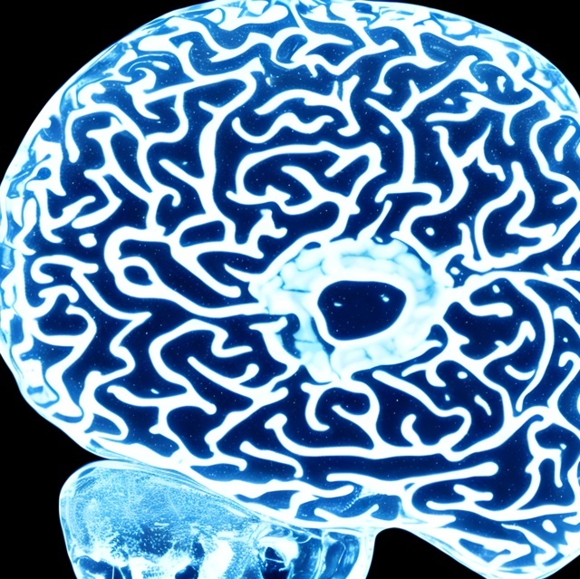
Hello, I would like to share with you some information about a fascinating technique called CLARITY that was developed in 2013 at Harvard University. CLARITY is a method that allows scientists to make tissues transparent and to visualize their molecular structures and functions in three dimensions. This has many applications in biomedical research, especially in the study of the brain and other complex organs. In this blog post, I will explain how CLARITY works, what are its advantages and limitations, and what are some of the current and future developments in this field.
How Does CLARITY Work?
CLARITY is based on the idea of transforming tissues into hydrogels, which are networks of water-soluble polymers that can retain the shape and integrity of the original tissue. To do this, the tissue is first fixed with formaldehyde, which crosslinks the proteins and nucleic acids to each other and to the hydrogel monomers. Then, the tissue is infused with acrylamide and bisacrylamide, which polymerize under heat and light to form the hydrogel matrix. The resulting tissue-hydrogel hybrid is then placed in a chamber filled with a detergent solution, such as sodium dodecyl sulfate (SDS), and an electric current is applied. This causes the detergent to remove the lipids from the tissue, which are the main source of opacity and scattering. The lipids are replaced by water, making the tissue transparent and permeable to molecules. The proteins and nucleic acids remain intact and attached to the hydrogel, preserving their location and structure. The transparent tissue can then be stained with fluorescent dyes or antibodies that bind to specific molecules of interest, such as genes, proteins, or neurotransmitters. The stained tissue can then be imaged with a microscope or a laser scanner, revealing its three-dimensional architecture and function. ¹
What are the Advantages of CLARITY?
CLARITY has several advantages over traditional methods of tissue analysis, such as slicing, staining, and reconstructing. First, CLARITY allows the visualization of intact tissues without sectioning or distortion, which can introduce errors and artifacts. Second, CLARITY enables the detection of multiple molecules simultaneously within the same tissue sample, which can reveal their interactions and correlations. Third, CLARITY allows the removal and reapplication of different stains or antibodies without damaging the tissue, which can facilitate multiple rounds of imaging and analysis. Fourth, CLARITY can be applied to various types of tissues and organs, such as brain, heart, liver, kidney, lung, spleen, pancreas, testis, bone, skin, eye, ear, etc. Fifth, CLARITY can be combined with other techniques, such as optogenetics (the use of light to control cells), electrophysiology (the measurement of electrical activity), or gene editing (the modification of DNA sequences). ²
What are the Limitations of CLARITY?
CLARITY also has some limitations that need to be addressed or improved. First, CLARITY is a time-consuming and labor-intensive process that requires specialized equipment and expertise. It can take several weeks or months to clear and stain a large tissue sample. Second, CLARITY can cause some loss or degradation of molecules during the clearing process, especially if repeated cycles of staining and destaining are performed. This can affect the sensitivity and accuracy of detection. Third, CLARITY can introduce some artifacts or biases in the imaging results due to uneven clearing or staining across different regions or depths of the tissue. This can affect the quantification and interpretation of data. Fourth,
CLARITY can pose some safety and ethical issues due to the use of toxic chemicals (such as formaldehyde or acrylamide) or human tissues (such as brain samples). These issues need to be carefully considered and regulated by appropriate guidelines and protocols.
What are some current and future developments in CLARITY?
CLARITY is a rapidly evolving technique that has been modified and improved by various researchers since its inception in 2013. Some of these modifications include:
– Using different types of hydrogels (such as PEG or agarose) or detergents (such as SDS or Triton X-100) to optimize the clearing efficiency or compatibility with different tissues or stains.
– Using different types of imaging modalities (such as confocal microscopy or light-sheet microscopy) or analysis software (such as ClearMap or NeuroGPS) to enhance the resolution or automation of data acquisition or processing.
– Using different types of applications (such as disease models or drug screening) or questions (such as connectivity mapping or cell type identification) to explore new aspects or functions of tissues or organs.
– Using different types of organisms (such as mice or zebrafish) or samples (such as embryonic or postmortem tissues) to expand the scope or relevance of research.
Some examples of current research projects using CLARITY include:
– Mapping the neural circuits and gene expression patterns in the mouse brain, revealing the structure and function of different brain regions and cell types.
– Visualizing the development and regeneration of the zebrafish heart, showing the dynamics and interactions of cardiac cells and tissues.
– Comparing the molecular and cellular profiles of normal and diseased human brains, identifying the changes and mechanisms associated with Alzheimer’s disease, autism, or schizophrenia.
Some examples of future research directions using CLARITY include:
– Developing faster, cheaper, and safer methods of tissue clearing and staining, using novel materials or techniques such as light-activated or enzyme-mediated hydrogels or detergents.
– Developing higher-resolution, deeper-penetration, and multi-modal imaging systems, using advanced technologies such as super-resolution microscopy or multiphoton microscopy.
– Developing more comprehensive, integrative, and interactive data analysis platforms, using artificial intelligence or machine learning algorithms to extract, visualize, and interpret complex information from large datasets.
– Developing more translational, clinical, and therapeutic applications, using human tissues or organs to diagnose, treat, or prevent various diseases or disorders.
Conclusion
CLARITY is a revolutionary technique that allows scientists to see through tissues and organs and to explore their molecular structures and functions in three dimensions. CLARITY has many advantages over traditional methods of tissue analysis, but it also has some limitations that need to be overcome or improved. CLARITY is a rapidly evolving technique that has been modified and improved by various researchers since its inception in 2013. CLARITY has many current and future developments that aim to optimize its performance and expand its applications. CLARITY is a powerful tool that can advance our understanding of biology and medicine and open new horizons for research and innovation.
References
– Chung K., Deisseroth K. (2013). CLARITY for mapping the nervous system. Nature Methods 10(6): 508–513.
– Tomer R., Ye L., Hsueh B., Deisseroth K. (2014). Advanced CLARITY for rapid and high-resolution imaging of intact tissues. Nature Protocols 9(7): 1682–1697.
– Yang B., Treweek J.B., Kulkarni R.P., Deverman B.E., Chen C.K., Lubeck E., Shah S., Cai L., Gradinaru V. (2014). Single-cell phenotyping within transparent intact tissue through whole-body clearing. Cell 158(4): 945–958.
– Economo M.N., Viswanathan S., Tasic B., Bas E., Winnubst J., Menon V., Graybuck L.T., Nguyen T.N., Smith K.A., Yao Z., Wang L., Gerfen C.R., Chandrashekar J., Zeng H., Looger L.L. (2018). Distinct descending motor cortex pathways and their roles in movement. Nature 563(7732): 79–84.
– Pan Y.A., Choy M., Prober D.A., Schier A.F. (2013). Robo2 determines subtype-specific axonal projections of trigeminal sensory neurons. Development 140(5): 978–987.
– Grinberg L.T., Rubino P.A., Ferretti R.E.L., Nitrini R., Farfel J.M., Polichiso L., Gierga K., Heinsen H., Suemoto C.K. (2017). The dorsal raphe nucleus shows phospho-tau neurofibrillary changes before the transentorhinal region in Alzheimer’s disease. A precocious onset? Neuropathology and Applied Neurobiology 43(5): 393–406.
– CLARITY Protocol and Tissue Clearing Guide | Abcam. https://www.abcam.com/protocols/clarity-protocol
– What is the CLARITY Technique? – News-Medical.net. https://www.news-medical.net/life-sciences/What-is-the-CLARITY-Technique.aspx
– CLARITY Methodology – News-Medical.net. https://www.news-medical.net/life-sciences/CLARITY-Methodology.aspx.
Tags
Divi Meetup 2019, San Francisco
Related Articles
Unappreciated Greatness
Life and Legacy of Jahangir of the Mughal Empire. Jahangir ruled over one of the largest empires in human history during his lifetime, yet few people outside of South Asia have heard of him. I aim to shed light on the life and legacy of this remarkable figure,...
The Plague Doctor’s Diary
A Personal Account of the Turin Epidemic of 1656. I am writing this diary to record my experiences and observations as a plague doctor in Turin, the capital of the Duchy of Savoy, during the terrible epidemic that has afflicted this city and its surroundings since the...
The Timeless Beauty of Bustan
Unveiling the Secrets of Saadi Shirazi's Masterpiece.In the realm of Persian literature, few works have captured the essence of love, spirituality, and morality quite like Bustan (The Orchard) by Saadi Shirazi. This 13th-century masterpiece has left a lasting impact...
Stay Up to Date With The Latest News & Updates
Explore
Browse your topics of interest using our keyword list.
Join Our Newsletter
Sign-up to get an overview of our recent articles handpicked by our editors.
Follow Us
Follow our social media accounts to get instant notifications about our newly published articles.


